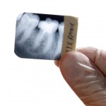
This review was undertaken to evaluate the diagnostic accuracy of available radiographic methods in use for imaging the periapical bone tissue area. The review forms part of wider systematic review covering methods of diagnosis and treatment in endodontics published by the Swedish Council on Health Technology Assessment (SBU). An English version of this is available on their website.
Searches were undertaken in the PubMed, Embase and the Cochrane Central Register of Controlled Trials (CENTRAL) databases. Two reviewers independently assessed abstracts and full text articles. An article was read in full text if at least one of the two reviewers considered an abstract to be potentially relevant. The GRADE approach was used to assess the quality of evidence of each radiographic method based on studies of high or moderate quality.
Twenty-six studies were included none were of high quality; 11 were of moderate quality.
The authors concluded
There is insufficient evidence that the digital intraoral radiographic technique is diagnostically as accurate as the conventional film technique. The same applies to cone beam computed tomography (CBCT). No conclusions can be drawn regarding the accuracy of radiological examination in identifying various forms of periapical bone tissue changes or about the pulpal condition.
The authors comment that at present, we have insufficient knowledge about the diagnostic accuracy of the different radiographic techniques in clinical use. They also highlight that a significant issue in evaluation of radiographic methods of periapical bone is that is that the reference test (gold standard) is either post-mortem study or biopsy!
Petersson A, Axelsson S, Davidson T, Frisk F, Hakeberg M, Kvist T, Norlund A, Mejàre I, Portenier I, Sandberg H, Tranaeus S, Bergenholtz G. Radiological diagnosis of periapical bone tissue lesions in endodontics: a systematic review. Int Endod J. 2012 Mar 19. doi: 10.1111/j.1365-2591.2012.02034.x. [Epub ahead of print] PubMed PMID: 22429152.
