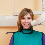
There have been a number of stories in the press regarding the recent paper by Claus et al on dental X-rays and meningiomas. Meningiomas are mostly benign tumours which arise from the dura mater and are usually slow-growing. They are the most common benign brain tumour although relatively uncommon with an incidence of around 6 per 100,000 and a female:male ratio is 2:1 . The aim of the Claus study was to examine the association between dental x-rays and the risk of intracranial meningioma.
What did they do?
The authors identified all individuals aged 20-70) with histologically confirmed intracranial meningioma in several states ( cases)over a 5 year period using Rapid Case Ascertainment (RCA) systems and state cancer registries. Controls were matched to cases by 5-year age interval, sex, and state of residence excluding those with previous history of meningioma and/or a brain lesion of unknown outcome.
Following approvals participants were contacted for a telephone interview, questions included onset, frequency, and type of dental care received including the number of times they had received radiographs (bitewing, full-mouth, or panoramic [panorex]), during 4 time periods: when aged <10 years, 10-19 yrs,20 -49 years, ≥50 years. Information also was gathered on the occurrence and timing of therapeutic radiation treatments.
A conditional logistic regression was used to assess the odds of meningioma associated with risk factors. individuals who had received therapeutic radiation were removed from all analyses that assessed the risk associated with dental x-rays
What did they report?
- Cases were more than twice as likely as controls to report having ever had a bitewings
- individuals who reported receiving bitewings on a yearly basis or more had an elevated risk for ages <
- An increased risk of meningioma also was associated with panorex films taken at a young age or on a yearly basis or more.
- No association was appreciated for tumour location above or below the tentorium.
[table id=25 /]
[table id=26 /]
What did they conclude
Our findings suggest that dental x-rays, particularly when obtained frequently and at a young age, may be associated with an increased risk of intracranial meningioma, at least for the dosing received by our study participants.
Claus, E. B., Calvocoressi, L., Bondy, M. L., Schildkraut, J. M., Wiemels, J. L. and Wrensch, M. (2012), Dental x-rays and risk of meningioma. Cancer. doi: 10.1002/cncr.26625
Things to consider
- This was a large study but there is no formal power calculation.
- The average length of the telephone interview was 52 minutes – How effective are telephone interviews at eliciting this type of information , is recall and honesty a greater or lesser problem than with a face to face interview?
- There is some discussion regarding the potential recall bias relating to the number and type of dental radiographs. This is discussed in the paper and it is suggested that recall for dental radiographs high but the studies quoted were much smaller.
- The controls were more likely to have ≥16 years of education and to have an annual salary >$75,000.
- It is interesting that ORs are higher for ‘bitewings at any age’ when you would anticipate that for older patients with longer dental histories there may potentially be a dose related effect so you might expect the ORs to be greater in the >50s.
- While the panorex data does suggest a higher risk these are all based on small numbers of patients.
- As noted in the Daily telegraph article, this is a rare disease so if this evidence was confirmed it would mean that the lifetime risk of meningioma would increase from 15 in every 10,000 people to 22 in 10,000
- Finally while this study does not raise significant concerns it is a timely reminder that dental x-rays should only be prescribed where there is a clear clinical need so as to reduce unnecessary exposure to ionising radiation.
Selection criteria for Dental radiographs from the Faculty of General Dental Practitioners
American Dental Association- use of dental radiographs
Further information on Meningiomas
Wiemels J, Wrensch M, Claus EB. Epidemiology and etiology of meningioma. J Neurooncol. 2010 Sep;99(3):307-14. Epub 2010 Sep 7. Review. PubMed PMID: 20821343; PubMed Central PMCID: PMC2945461.
http://www.patient.co.uk/doctor/Meningiomas.htm
http://www.macmillan.org.uk/Cancerinformation/Cancertypes/Brain/Typesofbraintumours/Meningioma.aspx

Huge problem, in that the control group is assumed to be disease free from a telephone interview. There is no evidence that they are disease free. There is only a lack of report of symptoms from a telephone interview.
The conclusion uses the word “dosing”. All we have is a person,s heavily biased recall. We have their memory of a childhood event. Not evidence of the radiographic dose.
And, of course we want to reduce radiographic exposure.
For further comment on this paper you could read the NHS Choices story X-ray ‘brain tumout risk’ not proven
Looked at your link, saw this “•More people in the case group reported having panoramic dental X-rays at a young age, on a yearly basis or with greater frequency compared with controls. For instance, individuals in the case group (with brain tumour) were almost five times more likely to report having received panoramic X-rays before the age of 10 than people in the control group (OR 4.9 95% CI 1.8 to 13.2).” That is almost 5 times the risk in this young group of subjects. The confidence interval is wide, not sure how that impacts the conclusion of the greater risk. Anyone want to comment on the interpretation of this CI?
individuals in the case group (with brain tumour) were almost five times more likely to report having received panoramic X-rays before the age of 10 than people in the control group (OR 4.9 95% CI 1.8 to 13.2).
This CI is derived from just 22 cases and 5 controls aged under 10 years of age. Is is a small number of patients which explains the wide CI but also raises concerns about whether there are enough patients to higlight this one finding so prominently in the abstract.
This study was completed by faculty at Yale University Sch. of Medicine. Principal investigator was Elizabeth B. Claus, MD, PhD, Department of Epidemiology and Public Health, Yale University School of Medicine, New Haven, CT. This person is a physician and a PhD. Others on the research group are highly qualified to design a valid research study. The real issue we should be discussing is that people should not be exposed to ionizing radiation inappropriately. I know many offices “take dental xrays according to how often the insurance will pay.” and not after a clinical exam reveals an indication. That is what we should be talking about.
I would agree that dental radiographs should only be taken when clinically justified my orginal blog had links to some well established guidance. However, despite the pedigree of the authors of the original authos and the journal itself there are still valid concerns regarding the study as the recent ADA statement higlights
This is indeed a very interesting study: I’ve been reading about it on the cancer.org website, as well as checking out the headlines and media reporting of this study.
This is the link to the cancer.org article:
http://www.cancer.org/Cancer/news/News/study-examines-possible-link-between-dental-x-rays-and-meningioma-risk
First and foremost, it goes without saying that we all need to approach dental radiography understanding that we are directing ionising radiation into another human’s body. The diagnostic benefits must always outweigh the risk, and we must do all we can to reduce dose (including investing in digital systems if we haven’t already done so, as well as ensuring that our clinical techniques are of excellent standard so that we always produce high-quality images and reduce the need for retakes, and ensuring that the appropriate shielding is provided for the subject of the radiographic exam and everyone else in the building).
The results of the study are concerning, and none of us like the idea that anything we do as dental practitioners might cause harm to others. But we also dislike hearing this sort of information because it potentially puts us into a defensive position: we find we have to justify what we do every day to people who are understandably concerned (even frightened) by this information and who themselves haven’t read the research or who may not completely understand the conclusions.
Dr Claus’ conclusion was:
“Exposure to some dental x-rays performed in the past, when radiation exposure was greater than in the current era, appears to be associated with an increased risk of intracranial meningioma. As with all sources of artificial ionizing radiation, considered use of this modifiable risk factor may be of benefit to patients.”
http://onlinelibrary.wiley.com/doi/10.1002/cncr.26625/abstract
Bottom line: that’s good advice, and I think (hope) that it (“considered use of this modifiable risk factor”) is the approach taken by many of our dental colleagues.
As dentists we need to be aware of the publication of this type of study (and the potential media frenzy that sometimes occurs as a result). We should read the study or at least the abstract. Then we can be knowledgable when we are asked questions about it. It also gives us an opportunity as clinicians and team leaders to consider our own approach to the practice of dentistry and make any positive modifications that may be necessary.
[…] […]