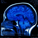
How brain imaging can help us understand mental health problems and their treatment is another service that the mental elf team will hopefully be able to offer. By blogging about interesting new studies we hope to help you pick up a wee bit of how MRI, CT, EEG and MEG adds to our understanding. While finding out that people who have different experiences of life, or symptoms of mental illnesses, have different brains is not surprising, the detail of what these differences are offers crucial information. It can provide clues as to the possible underlying causes and, in the future, allow for clearer diagnostic tests or predictions of what treatments will work.
Functional Magnetic Resonance Imaging (fMRI) allows researchers to estimate relative changes in brain activity. Blood changes colour based on how much oxygen there is in it: arteries are bright red, while veins are blue and MRI scanners can also pick up this change (Pauling, 1936). As a specific part of the brain is more active, more oxygen is used and the blood changes colour. From this researchers can work out which parts of the brain are more or less active when changing between different tasks (including doing nothing). This method does not capture all the activity in the brain associated with the task and there is ongoing debate about what stage of neuronal signalling is being imaged with fMRI.
A recent paper by researchers from London uses fMRI to try and better understand ADHD (Hart, 2013). They combined studies where people did specific tasks as a meta-analysis and found that the pattern of brain activity associated with both attention and inhibition is different in those people who have a diagnosis, compared to controls. They looked at the effect drug treatment might have and found that in one area of the brain, the activity was normalised by the medication.
Methods

People with ADHD usually find inhibition and attention tasks quite difficult
This meta-analytic study looked at the pattern of brain activity associated with two types of tasks:
- Inhibition: tasks where a person tries to stop themselves responding to a stimulus, despite learning that previously they should respond
- Attention: tasks where a person has to concentrate on specific stimuli
People with ADHD have consistently found these specific tasks hard, and a number of small fMRI studies have found that the pattern of brain activity whilst doing the tasks is different. The authors also looked for studies that measured the effect of taking stimulants, the mainstay of drug treatment for ADHD.
The authors found 21 data sets examining the inhibition tasks and 14 for attention. These covered both children (25 data sets) and adults (9 data sets) and totalled 287 people with ADHD and 320 people without. The authors did a good job of finding the relevant papers for inclusion but combining these types of studies is hard. Different scanners work slightly differently and comparing the images produced has been a challenge. It is a bit like taking lots of videos of a busy town square, using lots of different cameras, in different light and weather conditions and then trying to make an single clip of where, on average, people congregate. Great strides have been made in accounting for this variation but it still provides a real challenge for these types of studies. This paper was very conservative with its statistics. That means they may have missed some important findings, but we can be reasonably confident that the results are changes due to the tasks and ADHD, rather than methodological issues.
Results
- Areas of the brain associated with inhibiting movement were underactivated in people with ADHD
- Areas of the brain sensitive to where important stimuli are were also mostly underactivated
- In one area of the brain, the right caudate, treatment with stimulants made patients look like controls whilst performing attention tasks
Conclusions

This study does not mean that fMRI can be used to diagnose ADHD but will lead to further research on what might be underlying the changes seen
The authors conclude:
Patients with ADHD have consistent functional abnormalities in 2 distinct domain-dissociated right hemispheric fronto-basal ganglia networks [and that] preliminary evidence suggests that long term stimulant use maybe associated with more normal activation in the right caudate.
Providing a touch of elfin translation to the authors conclusions:
- The pattern of brain activity associated with the difficulties people with ADHD experience is becoming clearer
- There is a general reduction in activity across specific brain networks related to attention and inhibition
- In one key hub of the network, treatment normalises the brain activity, but in all the other areas it does not
This study does not mean that fMRI can be used to diagnose ADHD but will lead to further research on what might be underlying the changes seen. The effect of medication on one part of the brain, rather than elsewhere, again offers clues as to what might be going on to make the drugs work, but further research will be needed to work out how useful these scanning findings are to day to day clinical practice.
Links
Hart H, Radua J, Nakao T, Mataix-Cols D, Rubia K. Meta-analysis of fMRI studies of inhibition and attention in attention deficit/hyperactivity disorder: exploring task-specific, stimulant and age effects. JAMA Psychiatry. 2013 70(2):185-98 [Abstract]
Pauling L, Coryell CD. The Magnetic Properties and structure of hemoglobin, oxyhemoglobin and carbonmonoxy hemoglobin (PDF). Proc. Nayl. Acad. Sci.(USA). 1936 22:210-16


We’ve started a new series of brain imaging blogs written by @andrewwatson28 The first one is about #ADHD http://t.co/N3Yy5prqJe
great blog demystifying scanning research into adhd. thanks @Mental_Elf ! http://t.co/MhAX9zhVIP
@Mental_Elf what is ‘normal’? They should quit messin with people & let them be themselves instead of tryin make them ther version of normal
New meta-analysis uses Functional Magnetic Resonance Imaging to measure inhibition & attention in #ADHD http://t.co/N3Yy5prqJe #fMRI
The pattern of brain activity associated with the difficulties ppl with ADHD experience is becoming clearer http://t.co/N3Yy5prqJe
@Mental_Elf yet it’s already being treated with drugs “thought to…”
There is a general reduction in activity across specific brain networks related to attention and inhibition http://t.co/N3Yy5prqJe
Read the first of our brain imaging blogs by @andrewwatson28 Clinical Research Fellow at Edinburgh University http://t.co/N3Yy5prqJe
great job. thanks. hope you do more of this. does brain behaviour in GAD, anxiety show overactive amygdala system?