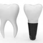
The main aim of this systematic review is to look at implant based restorative options for restoring the atrophic posterior mandible using osseointegrated dental implants. The atrophic posterior mandible is defined here as having a residual ridge height of 8mm from the inferior dental nerve to the crest of the ridge. The two options considered are short dental implants < 8mm in length placed in pristine bone compared to standard long implants >8 mm placed after bone augmentation procedures, in this care either autogenous onlay grafts or non-onlay autogenous and xenogeneic bone grafts
Methods
This review followed the PRISMA statement. Searches were screened by two independent researchers using Medline, and Cochrane Oral Health Group databases. Databases were searched from January 1st 2006 to July 30th 2016 and restricted to English, manual searches were also carried out in the relevant major journals.
Inclusion criteria were: Randomised clinical studies (RCT’s) that included clinical or radiological outcomes of the surgical strategies for rehabilitation of atrophic posterior mandibles in partially edentulous patients. This including any dimensional change, survival rate and adverse event and follow-up from 12 to 24 months. Excluded studies included animal studies, repeated reports of the same study, and studies including patients who were heavy smokers, drinkers, or had poor oral hygiene.
Quality appraisal was carried out by each of the authors using the Cochrane Collaboration tool for assessing risk of bias in randomised trials. The primary outcomes were implant failure and marginal bone loss. Secondary outcomes were biological complications and prosthesis failure.
Results
- From 138 records only 12 fulfilled the inclusion criteria. A total of 353 patients with 674 implants were treated.
- None of the papers selected were judged as having a low risk of bias.
- The authors ranked the studies into three categories:
- Group A: 5 studies which compared outcomes of standard implants placed in augmented bone (long implants group) vs. outcomes of short implants placed in pristine bone (short implants group).
- Group B: 4 studies which compared outcomes of standard implants placed in bone augmented with onlay block (onlay blocks group) vs. outcomes of standard implants placed in augmented bone with any of the other augmentation procedures that did not involve onlay blocks (non-onlay blocks group).
- Group C: 4 studies not included in category A) or B). Meta-analysis could not be performed.
| Group A | Risk Ratio | 95% Confidence Interval | P-Value |
| Short implant v. long implant (Implant failure) | 1.59 | 0.54 to 4.69 | 0.397 |
| Short implant v. long implant (Prosthesis failure) | 1.49 | 0.0.56 to 3.96 | 0.426 |
| Short implant v. long implant (biological complications) | 2.82 | 1.81 to 4.4 | <0.0001 |
| Mean Diff | 95%Confidence Interval | P-Value | |
| Short implant v. long implant (marginal bone loss) | 0.05 mm | 0.026 to 0.079 | <0.0001 |
| Group B | Risk Ratio | 95% Confidence Interval | P-Value |
| Onlay Group v. Non-onlay Group | 1.81 | 0.42 to 7.84 | 0.43 |
| Mean Diff | 95% Confidence Interval | P-Value | |
| Onlay Group v. Non-onlay Group | 0.006 mm | -0.19 to 0.177 | 0.946 |
Conclusions
The authors concluded:-
Findings from subgroup analyses revealed that
(1) short implants placed in the posterior atrophic areas of partially edentulous mandibles were associated with superior outcomes compared with long implants in augmented bone, such as lower rate of biological complications and of peri-implantbone loss; whereas
(2), there was no evidence that onlay augmentation was inferior to any of theother augmentation techniques employed
Comments
Even though the results of this systematic review and meta-analysis favour short implants over standard implants placed in augmented bone the evidence must be interpreted with extreme caution for the following reasons:
- A wider literature search may have found more relevant papers.
- There were no studies that were at a low risk of bias.
- The mean number of implant in the studies were 28/arm of study leading wide confidence intervals
- The study durations were very short and only one study extended beyond 4 years, 5 out of 12 studies were only 12 months long.
In general, there are issues with statistical v. clinical significance. Even though the risk ratio favours short implants in terms of reduced biological complications there are no other metrics where there is a visible clinical difference; an example would be marginal bone loss of 0.05mm. Some qualitative research would be interesting to explore the patient’s experience of bone augmentation procedures in terms of morbidity against short implant placement into pristine bone.
Links
Primary paper
Toti, P. et al., 2017. Surgical techniques used in the rehabilitation of partially edentulous patients with atrophic posterior mandibles: A systematic review and meta-analysis of randomized controlled clinical trials. Journal of Cranio-Maxillofacial Surgery.
Other references
Dental Elf – 2nd Dec 2015
Dental Elf – 3rd June 2016

Very good Mark. Short implants are always an option. However, clinically our concerns remain around bone loss in an ever growing problem
Ever growing in Peri-implantitis cases where 1-2mm of bone loss on a 5-6mm length implant means even less bone holding the implant in. Generally these short implants have large molar teeth on them also.
Hi TC
Thanks for the input. Agreed, on the positive side at least short implants with peri-implantitis are easier to get out than those 12+mm in the aesthetic zone ;)
[…] Short dental implants for the atrophic posterior mandible? […]