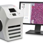
Digital pathology encompasses the same principles of histological assessment with conventional light microscopy whereby a surgical pathology specimen is processed, stained and mounted on a glass slide. The digital component of the pathway involves a robotic microscope that performs whole slide imaging (WSI) where sections of the slide are scanned at different focus distances to allow for a greater depth of field and assessment at several magnifications.1 Once the images have been captured, they are digitally assembled to produce a complete virtual slide, viewed on a digital screen by a pathologist.2 The authors aimed to conduct a systematic review and meta-analysis to review evidence across validation studies and summarise the safety and reliability of digital pathology.
Methods
The Preferred Reporting Items for Systematic Review and Meta-Analyses were used to inform the methodology of this review. PubMed, Medline, EMBASE, Cochrane Library and Google Scholar were the databases searched. Additionally, manual searches were conducted via forward citation tracking and reference searches of included studies. Quality assessment was undertaken through the Quality Assessment of Diagnostic Accuracy Studies (QUADAS 2) tool.
Results
- 994 articles were retrieved across the initial database and manual search. Following removal of duplicates and implementation of the inclusion and exclusion criteria, 24 studies were included in this review.
- 23 studies were conducted at single centres and 2 were multi-centre validations. 14 studies were from America, 10 from Europe and 1 from Asia.
- 56% of studies were published from 2015 onwards.
- The sample sizes varied from 60 to 3017 cases across a range of specialities. The length of washout time between digital pathology and light microscopy reporting ranged from 2 weeks to more than a year. 4 studies did not use a washout time due to a live-validation approach.
- Overall concordance was assessed by measuring agreement along with clinically insignificant variations between digital and light microscopy reports. The pooled agreement was 98.3% (95% CI 97.4-98.9).
- 546 major discordances were reported across 21 studies with gastrointestinal tract and gynaecological pathology the most commonly reported specialities with these. 57% of discordances were related to the assessment of nuclear atypia, grading of dysplasia and malignancy.
- A low risk of bias was reported in 75%-92% studies and high risk of bias in 8-25% across all QUADAS 2 domains.
Conclusions
The authors concluded:-
…The results of this meta-analysis represent significant evidence to indicate the equivalent performance of digital pathology for routine diagnosis.
Comments
The authors have conducted a robust and the largest systematic review and meta-analysis to date of digital pathology. The high percentage concordance between digital pathology and light microscopy provides further evidence that digital pathology is a viable alternative to light microscopy. The authors acknowledged the limitations of this review in that included studies had no ophthalmic samples and few renal or paediatric samples for conclusions to be drawn on these. Therefore, while this review highlights good concordance, specific areas for future research in digital pathology are highlighted. With regards to head and neck pathology, seven studies assessed this discipline but no specific findings reported. There is a momentum shift towards digital pathology, likely accelerated by the emphasis of home-working and maintaining service provisions during the COVID-19 pandemic. The necessary Information Technology infrastructures need to be integrated into current reporting work streams and digital pathology training included in the specialty training curriculum to help with the near future transition to digital pathology.
Links
Primary Paper
Azam AS, Miligy IM, Kimani PK, et al (2020). Diagnostic concordance and discordance in digital pathology: a systematic review and meta-analysis. Journal of Clinical Pathology doi: 10.1136/jclinpath-2020-206764
Other references
- Zarella MD, Bowman D, Aeffner F, Farahani N, Xthona A, Absar SF, et al. A Practical Guide to Whole Slide Imaging: A White Paper From the Digital Pathology Association. Archives of pathology & laboratory medicine. 2019;143(2):222-34.
- Indu M, Rathy R, Binu MP. “Slide less pathology”: Fairy tale or reality? Journal of oral and maxillofacial pathology : JOMFP. 2016;20(2):284-8.
Picture Credits
www.leicabiosystems.com, CC BY-SA 4.0, via Wikimedia Commons
