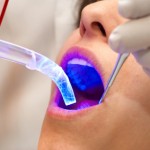
Worldwide 263,000 new cases or oral cancer are seen each year with around 127,700 deaths. In the UK the incidence has increased since the 1980s. As outcomes are better with early detection approaches to improve on clinical examination alone have been developed. The aim of this systematic review is to evaluate the literature investigating the effectiveness of chemiluminescence and autofluorescent imaging devices as aids in the detection of oral squamous cell carcinomas (OSCC) and oral potentially malignant disorders (OPMD) and for formulating recommendations for their use in primary care settings and by specialists.
Methods
A search was conducted in the Medline/PubMed database for primary studies where an optical device was used for investigation, screening or as a diagnostic tool for OSCC or OPMD. Data was abstracted independently by two reviewers
Results
- 25 studies were included ,13 used chemiluminescence, 12 used tissue autofluorescence.
- Biological plausibility, technical feasibility and diagnostic performance of the various systems are considered
- Chemiluminescence shows good sensitivity at detecting any OPMDs and oral cancer. However, it preferentially detects leukoplakia and may fail to spot red patches. The additive use of toluidine blue may improve specificity.
- Tissue autofluorescence is sensitive at detecting white, red and white and red patches, and the area of fluorescence visualisation loss (FVL) often extends beyond the clinically visible lesion. However, in addition to OPMDs, VELScope may detect erythematous lesions of benign inflammation resulting in false-positive test results.
Conclusions
The authors concluded:
There is limited evidence for their use in primary care, and these tools are better suited to specialist clinics in which there is a higher prevalence of disease and where experienced clinicians may better discriminate between benign and malignant lesions.
Commentary
Limiting the number of databases searched and studies to those in English only raises the possibility that some relevant studies may have been excluded. Although the authors do note two Spanish reviews with similar findings. While no data aggregation has been conducted the included studies are discussed in detail. The authors highlight the challenges with the definition of leukoplakia, which was variable across the studies. They also note that in primary care,
conventional visual inspection under normal incandescent light, followed by biopsy of suspicious lesions, will remain the gold standard for the immediate future.
The authors also highlight that neither chemiluminescence or autofluorescence improve on the level of sensitivity and specificity achieved by conventional oral examination (Walsh et al 2013).
Links
Rashid A, Warnakulasuriya S. The use of light-based (optical) detection systems as adjuncts in the detection of oral cancer and oral potentially malignant disorders: a systematic review. J Oral Pathol Med. 2014 Sep 3. doi: 10.1111/jop.12218. [Epub ahead of print] PubMed PMID: 25183259.
Brocklehurst P, Kujan O, O’Malley LA, Ogden G, Shepherd S, Glenny AM. Screening programmes for the early detection and prevention of oral cancer. Cochrane Database of Systematic Reviews 2013, Issue 11. Art. No.: CD004150. DOI: 10.1002/14651858.CD004150.pub4. – See more at: http://www.thedentalelf.net/2013/11/26/limited-evidence-to-decide-whether-visual-screening-reduces-the-death-rate-for-oral-cancer/#sthash.co4wU8bF.dpuf
Walsh T, Liu JLY, Brocklehurst P, Glenny AM, Lingen M, Kerr AR, Ogden G, Warnakulasuriya S, Scully C. Clinical assessment to screen for the detection of oral cavity cancer and potentially malignant disorders in apparently healthy adults. Cochrane Database of Systematic Reviews 2013, Issue 11. Art. No.: CD010173. DOI: 10.1002/14651858.CD010173.pub2.

“@TheDentalElf: Oral cancer detection: http://t.co/mKtxYfZoaj” MT best method currently is visiting your dentist regularly
@MangerBakes @Toothfairy4you @TheDentalElf en mondhygiëniste :-)