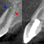
Root development in immature tooth requires a health dental pulp. Trauma or infection may result in pulp necrosis stopping further tooth development leaving open apical foramina, large root canals, and thin canal walls. Typical in these circumstances apexification using and mineral trioxide aggregate (MTA) is the main treatment choice however it does not allow further root development potentially compromising the tooth. The re-establishment of a functional pulp is necessary to promote continued root formation with successful pulp revascularisation being first reported in 2001. Revascularisation procedures involve root canal disinfection and blood clot induction with favourable and favourable outcomes being reported. The apical diameter is considered to be an important factor in achieving a favourable outcome.
The aim of this review was to evaluate whether the apical diameter of teeth with necrotic pulp affects the outcomes of regenerative endodontic treatment ( RET) and to determine the minimal apical size needed to obtain proper pulp revascularization.
Methods
Searches were conducted in the Cochrane Central register of controlled trials, Medline, Embase and clinical trials.gov databases supplemented by hand searching of the journals, Australian Endodontic Journal; Dental Traumatology; International Endodontic Journal; Journal of Endodontics; and Oral Surgery, Oral Medicine, Oral Pathology, Oral Radiology and Endodontology. Studies reporting teeth diagnosed with pulp necrosis treated by RET in humans with a minimum of 3 months follow up were considered. The primary outcomes were, Clinical or radiographic signs and symptoms of periapical healing and radiographic signs of continued root development.
Two reviewers independently selected studies and abstracted data and the Newcastle-Ottawa Scale was used to assesses methodological quality and a narrative summary of the findings presented.
Results
- 14 studies (1 prospective cohort study, 2 prospective case series, 1 retrospective case series and 10 case reports) were included.
- Apical diameters were measures using a range of different approaches.
- 95 teeth were separated into 3 groups based on apical diameter and reported in paper as outlined in table.
| Apical diameter | No. teeth | Healed | Incomplete
Healing |
Failed | Excluded | Clinical Success |
| Narrow <0.5 mm | 10 | 2 | 7 | 1 | – | 90.0% |
| Medium 0.5–1.0 mm | 25 | – | – | 1 | 2 | 95.7% |
| Wide >1.0 mm | 60 | – | – | 4 | 7 | 92.9% |
Conclusions
The authors concluded: –
Within the limitations, the review suggests that teeth with an apical diameter <1.0 mm achieved clinical success after RET (ie, 90% in group N [<0.5 mm], 95.65% in group M [0.5–1.0 mm], and 92.98% in group W [>1.0 mm]). The teeth with apical diameters of 0.5–1.0 mm attained the highest clinical success rate, which may be related to other potential factors, including patient age, pulp necrosis aetiology, preoperative apical radiolucency, procedure details, follow- up period, or sample size. Thus, the conclusion should be interpreted with caution.
Comments
The reviewers have undertaken an extensive search for relevant studies. However, the available studies are only small case series and reports involving small number of patients. The authors used three categories to assess primary outcomes :-
- Complete healing: absence of clinical signs and symptoms, complete resolution of peri-radicular radiolucency, and increased root dentin thickness/length and apical closure
- Incomplete healing: absence of clinical signs and symptoms, reduced or unaltered periapical lesion size with/without radiographic signs of increasing root dentin thickness/length, or apical closure
- Failure: persistent clinical signs and symptoms and/or increased peri-radicular lesion size
Outcomes 1 and 2 were both considered clinical success and the authors present percentage clinical success rates for three apical diameters although unfortunately it is not clear from the data presented how these are achieved as figures for healed and incompletely health teeth are not clearly provided in the paper.
Given the low quality of the available evidence the findings should be treated very cautiously and while the data suggest that RET may be a potential treatment there is a clear need for high quality well conducted and reported studies to truly determine whether this is a viable approach. The availability of only low quality evidence for RET was also highlighted in a broader review of the topic we have looked at previously (Dental Elf – 18th Sep 2017 ).
Links
Primary Paper
Fang Y, Wang X, Zhu J, Su C, Yang Y, Meng L. Influence of Apical Diameter on the Outcome of Regenerative Endodontic Treatment in Teeth with Pulp Necrosis: A Review. J Endod. 2018 Mar;44(3):414-431. doi: 10.1016/j.joen.2017.10.007. Epub 2017 Dec 19. Review. PubMed PMID: 29273495.
Original review protocol on PROSPERO
Other references
Dental Elf – 18th Sep 2017
Regenerative endodontic treatment or MTA apical plug in teeth with pulp necrosis and open apices?
Picture Credits
