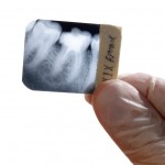
Although non-surgical root canal treatment has high long term success rates failures do occur. The literature indicates that poor coronal seal is associated with increased an increased failure rate with coronal microleakage occurring in days to week even in cases with apparent high quality root canal obturation. The placement of over canal restorations or orifice barriers has been suggested as a method of improving the coronal seal and a range of restorative materials have been tested. However prospective studies have only been conducted in animals with in-vitro studies conducted in human teeth under controlled conditions.
The aim of this review was to compare the efficiency of orifice barriers in preventing coronal microleakage in-vitro.
Methods
A review protocol was registered on the PROSPERO database. Searches were conducted in the Medline/PubMed, Embase, Scopus, Wanfang Database and China National Knowledge Infrastructure (CNKI) and Web of Science databases. in vitro studies comparing microleakage of root canal treated extracted human teeth with and without placement of an orifice barrier material published in English, German and Chinese were considered. Two reviewers independently selected studies and extracted data. Risk of bias was assessed using the QUIN tool (Sheth et al, 2022). Data was assessed qualitatively and quantitatively. Risk ratios (RR) and 95% confidence intervals (CI) were presented, and meta-analyses were conducted.
Results
- 18 in-vitro studies were included using a total of 33 different orifice barriers.
- The most frequently used materials were glass ionomer cement (GIC), resin modified GIC (RMGIC), composite resin, mineral trioxide aggregate (MTA) and Cavit.
- 15 studies evaluated microleakage by turbidity of the bacterial growth medium while 3 studies used a fluid filtration model.
- Meta-analysis was possible for GIC, RMGIC, Composite resin, MTA and Cavit tested with a bacterial leakage model.
- The table below shows risk ratios for a range of materials compared to no barrier placement.
| Material | No. of studies | Risk ratio (95%CI) |
| Glass ionomer cement (GIC) | 4 | 0.37 (0.26 to 0.53) |
| Resin modified GIC (RMGIC) | 6 | 0.32 (0.15 to 0.67) |
| Composite resin (CR) | 2 | 0.54 (0.38 to 0.75) |
| Mineral trioxide aggregate (MTA) | 3 | 0.25 (0.12 to 0.52) |
| Cavit | 2 | 0.23 (0.14 to 0.39) |
- Pairwise comparisons between a number of different materials was possible but no significant differences were demonstrated (see table below).
| Comparison | No. of studies | Risk ratio (95%CI) |
| GIC v CR | 2 | 0.50 (0.25 to 1.02) |
| GIC v RMGIC | 2 | 1.89 (0.10 to 37.13) |
| RMGIC v MTA | 3 | 1.12 (0.56 to 2.22) |
Conclusions
The authors concluded: –
Placement of an orifice barrier over the root canal filling is effective in the prevention of coronal microleakage in vitro. Other parameters may also affect the effectiveness of orifice barriers, including thickness and duration of exposure to the oral environment.
Comments
This review of in-vitro studies assessing the efficacy of orifice barriers included 18 studies all of which were considered to be at medium risk of bias. Unsurprisingly all the materials tested performed better than the control of no barrier. Two-way comparisons were performed for a small number of materials both these involved small numbers of studies (2 -3) with no statistically significant differences between the materials tested. When considering the findings of this review it needs to be remembers that all the studies were conducted in-vitro which may not be fully representative of the in-vivo situation. Dye leakage studies were not included in this review as for a number of technical reasons may overestimate leakage and also correlate poorly with other methodologies. So while the findings suggest a benefit from an orifice barrier the findings should be viewed cautiously and balanced with the potential difficulty of its removal should the need arise.
Links
Primary Paper
Chen P, Chen Z, Teoh YY, Peters OA, Peters CI. Orifice barriers to prevent coronal microleakage after root canal treatment: systematic review and meta-analysis. Aust Dent J. 2023 Jun;68(2):78-91. doi: 10.1111/adj.12951. Epub 2023 Feb 9. PMID: 36661351.
Other references
Sheth VH, Shah NP, Jain R, Bhanushali N, Bhatnagar V. Development and validation of a risk-of-bias tool for assessing in vitro studies conducted in dentistry: The QUIN. J Prosthet Dent. 2022 Jun 22:S0022-3913(22)00345-6. doi: 10.1016/j.prosdent.2022.05.019. Epub ahead of print. PMID: 35752496.
Dental Elf – 29th Apr 2022
Endodontic sealers and the outcome of endodontically treated permanent teeth
